
2007) see Critical Parameters for guidance.

Images can be taken with a flatbed scanner or digital camera ( Dahle et al.
#CELLPROFILER OUTPUT DURING PROCESSING PATCH#
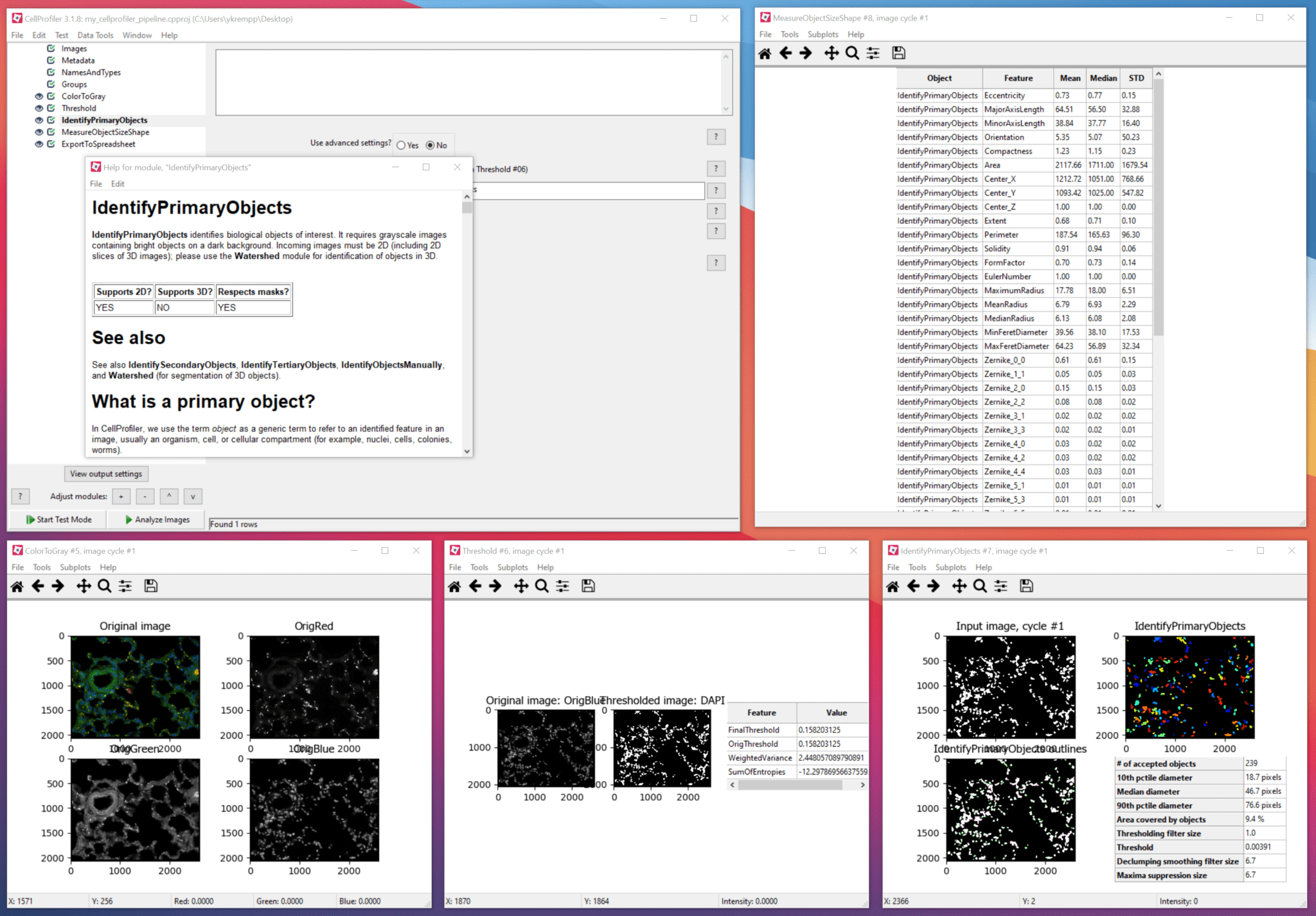
CellProfiler has been cited in more than a thousand papers and validated for a wide variety of biological applications, including yeast colony counting and classification, cell microarray annotation, yeast patch assays, cell-cycle classification, mouse tumor quantification, wound healing assays, and tissue topology measurement, as well as analysis of fluorescence microscopy images for measurement of cell size and morphology, cell cycle distributions, fluorescence staining levels, and other features of individual cells in images ( Lamprecht, Sabatini, and Carpenter 2007 Carpenter et al. The protocol uses the open-source, freely downloadable software package, CellProfiler. This unit outlines a protocol for the automated counting and analysis of yeast colonies growing on agar plates however, the methods described can be adapted to a wide variety of biological “objects” and can be used to measure a wide variety of features for each object. It is less tedious, more objective and quantitative, and, while the set up can be time-consuming, the analysis itself is usually much faster for large sample sets. Acquiring images and analyzing them automatically with image analysis software has several advantages over simple visual inspection. Many experiments in a biology laboratory involve visual inspection, such as examining yeast colonies or growth patches on agar plates, or examining live or stained cell samples by microscopy.


 0 kommentar(er)
